Once upon a time, William Woods students in the EXS 205 – Intro to Anatomy class did their learning the old fashioned way; that is, they studied 2D models in textbooks and PowerPoints. But this year, the students have begun learning while using new 3D anatomical models after Dr. Steve Middleton, Assistant Professor of Athletic Training and the Director of the Master of Science Athletic Training program, introduced an expanded amount of anatomical modes to the class.
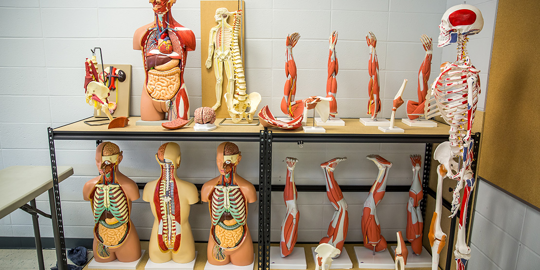
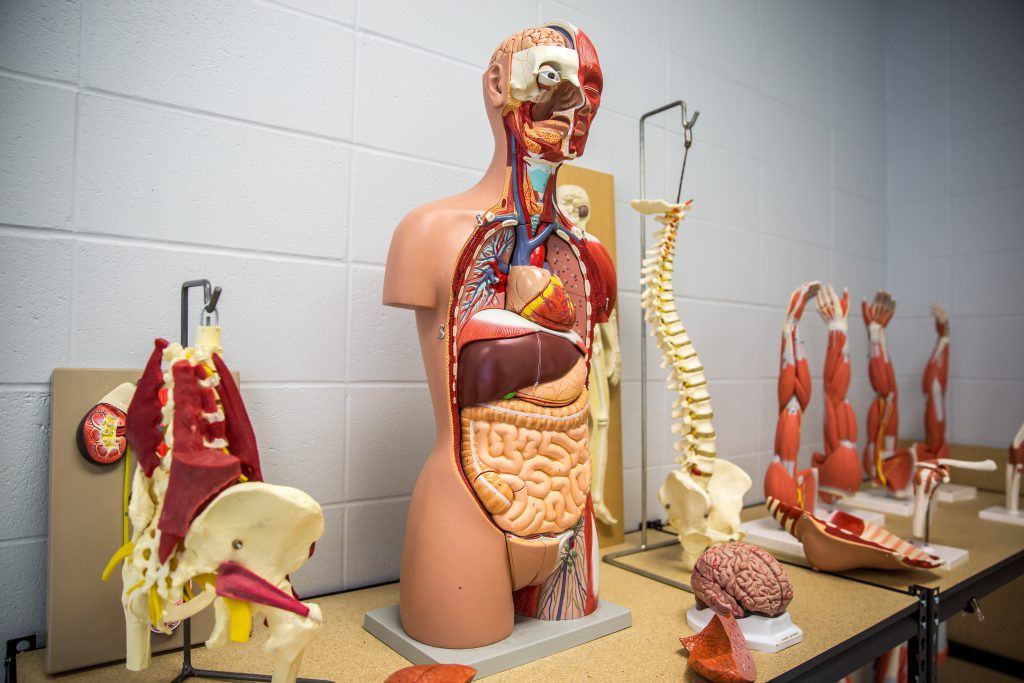
Upon Dr. Middleton’s arrival in Fall 2015, the anatomy course had a few small models of specific organs and some skeletal models that were in a state of disarray. One of Dr. Middleton’s first priorities was to propose a purchasing plan to establish a basic lab. The lab currently contains three muscled arms, three muscled legs, three torsos with removable organs, multiple skeletons, and advanced joint models that allow athletic training students to practice joint manipulations. There is now a plan in place to expand the lab with more models so that fewer students must share a single model. There may be additional specialty models added over time as well.
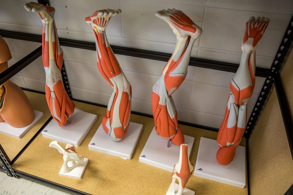
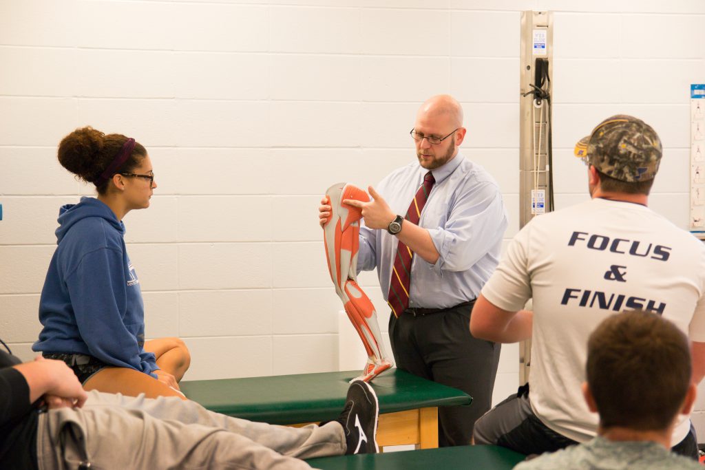
To prepare the students for their exams about identifying structures on the models ranging from bony landmark to specific muscles to organs, Dr. Middleton divides the class in two for one class period while the class goes over the leg model. For the next class period, the class was broken up again to learn about the arm.
“The class gives students a hands-on learning opportunity, which is one of the hallmarks of the university as a whole.” Middleton said.
When sending a survey to the EXS 205 Intro to Anatomy students, 21 of the 24 students responded. 19 of those students found the models useful for class. One of those students was Alexis Hassler ‘22, who is majoring in Exercise Science with a concentration in Athletic Training. Hassler believes both the models and PowerPoints help her with improving her knowledge for quizzes and tests.
“I like having the PowerPoints especially when they are uploaded to OwlNet to study because not everything can be seen on the models and there is other information we need to know that doesn’t involve the models,” Hassler said. “The models are great though, especially when we break into small groups and go over things using the models.”
With the addition of the models, Middleton visualizes the future of Into to Anatomy class. His goal is to have a cadaver lab, which is where someone has donated their body to science for students to learn the anatomy by dissecting one of those individuals.
“It is where I hope to go in the future especially as we start adding health care majors,” Middleton said. “While the textbooks are good and the models are good, there is nothing like learning from the body itself because there is a lot of anatomical variations from person to person. Dissection changes a person; not just the person being dissected but the person performing it as well. You start to appreciate how our lifestyles affect us.”
For the students, the class is one of the core classes for Exercise Science and fulfills one of the university’s general education requirement for natural sciences. The class is full every fall and every spring when it is offered. The department has just expanded the course offering from one section in the fall to two sections allowing an additional 20-24 students to take the course.
“We spend so much time learning about other things, but we do know about our own bodies and that we are made to work.” Dr. Middleton said. “We talk about structural changes associated with obesity, smoking, diets, and things of that nature. It helps students learn what we are doing to ourselves with our activities of daily living.”
And with the addition of anatomical models, William Woods students who study the sciences will have new resources to help them learn well into the future.